What is coarctation?
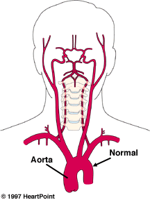
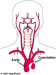
Coarctation of the aorta is a narrowing of the aorta, the main blood vessel carrying oxygen-rich blood from the left ventricle of the heart to all of the organs of the body.
Coarctation occurs characteristically in a short segment of the aorta just beyond where the arteries to the head and arms take off, as the aorta arches inferiorly toward the abdomen and legs.
Coarctation occurs in the "juxta-ductal" part of the aorta, or the part near where the ductus arteriosus attaches (Please see the illustration.)
Because the ductus arteriosus is a blood vessel that has special tissue in its wall that causes it to close in the first hours or days of life, some believe that coarctation is caused by the presence of extra ductal tissue extending into the adjacent aorta which results in aortic narrowing as the ductal tissue contracts.
Coarctation may be associated with other cardiac defects, typically also involving the left side of the heart. Most commonly seen are bicuspid aortic valve and ventricular septal defect. Coarctation may also be seen as a part of more complex, single ventricle cardiac defects.
Coarctation forces the left ventricle to work harder. It must generate a higher pressure than normal to force blood through the narrow segment of the aorta to the lower part of the body.
If the narrowing is severe, the ventricle may not be strong enough to perform this extra work resulting in congestive heart failure or poor perfusion to the organs of the body.
Detecting coarctation
The age at which coarctation is detected depends on the severity of the narrowing.
In approximately 50 percent of cases of isolated coarctation, the problem is severe enough to cause symptoms in the first days of life coinciding with the time that the ductus arteriosus closes. In these cases, infants may be noted to have signs and symptoms of severe congestive heart failure or shock.
Inability to feel pulses in the legs typically directs the physician's attention to the possibility of coarctation in such an ill infant.
Echocardiography is most commonly employed to define the aortic anatomy and evaluate for other cardiac anomalies.
When a coarctation goes undetected in the newborn period, it may go unrecognized for many years in some cases. The most frequent findings leading to its detection are a murmur, weak lower extremity pulses, or hypertension (high blood pressure) in the arms.
Echocardiography is the most common test used to confirm the diagnosis, but occasionally in older children this region is difficult to image well by echo. In such cases, MRI or CT scan may be employed.
Managing coarctation
In a critically ill newborn, ventricular function must be supported, often with the use of medications. Initiation of prostaglandin, which relaxes ductal tissue, may be employed in the initial stabilization.
Surgical repair is typically carried out on an urgent basis in symptomatic newborns with coarctation. While a number of surgical techniques may be employed, including patching or the use of an arterial (left subclavian artery) flap, resection (removal) of the area of narrowing and direct reanastomosis (reconnecting) is most commonly performed.
In older children, coarctation repair is typically planned electively if the diagnosis is made after the child is a year old.
While surgical resection and end-to-end reconstruction is performed most commonly, catheter-based therapy such as balloon dilation or balloon dilation with stent placement may be considered in selected cases.
Balloon dilation as initial therapy for coarctation is somewhat controversial, though it is widely accepted as the primary treatment for recurrent coarctation following surgical repair.
Dilation and stenting is a newer modality, offered to selected older children. Long-term follow-up studies will be needed to adequately assess this technique, though early results are encouraging.
Coarctation of the aorta treatment results
Potential complications of operations on the aorta are damage to downstream organs, particularly the kidneys or the spinal cord, during the time blood flow is interrupted for the repair.
In children with coarctation, though, these complications are very rare due to the presence of "collateral" or detour blood vessels that channel blood flow around the narrowing to the lower segments of the aorta.
Restenosis of the aorta at the area of the coarctation repair is possible, even years following treatment. The risk of restenosis is highest among newborns with rates of approximately 10 percent.
In older children the rates of restenosis after surgical repair of coarctation are much lower, approaching zero by age 3 years.
The chance for restenosis is higher after balloon dilation; between 20 percent and 25 percent. While operation is recommended in some patients for restenosis, the majority of cases are managed with balloon dilation as the initial approach.
Another concern after coarctation repair is high blood pressure. While this is rarely seen in infants, most older children have unusually high blood pressure requiring aggressive treatment immediately following surgery.
Long-term hypertension is also a concern in older children even after coarctation repair. The chances of having chronic high blood pressure problems requiring treatment are greater the older a child is at the time of repair. This is likely due to changes that occur in the blood vessels throughout the body in response to the presence of a coarctation.
Long term follow-up with the cardiologist is important for children after coarctation treatment to diagnose late problems of restenosis or hypertension.
Another reason continued follow-up is important is because approximately 30 percent of patients with coarctation also have a bicuspid aortic valve for which endocarditis prophylaxis should be followed.
Such a valve abnormality may lead to problems later in life. Follow-up visits often include a physical exam including blood pressure measurement in both the arms and the legs, periodic electrocardiograms and echocardiograms.


 my Cowboys